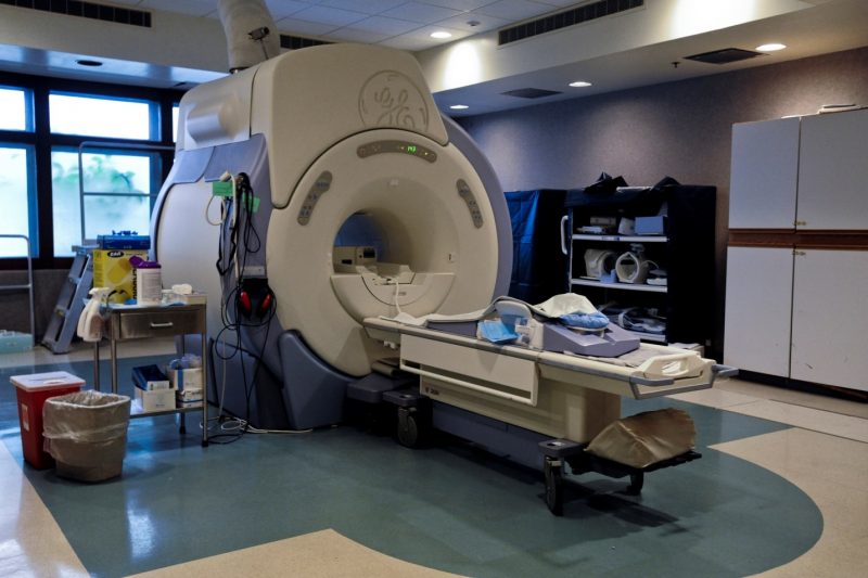
MRI (Magnetic resonance imaging) plays a revolutionary role in today’s world of medicine, as the most informative and versatile diagnostic imaging, especially in neurological procedures. According to Dr Timothy Steel, knowing what an intracranial lesion accurately first is a must in managing the lesion. The MRIs radio frequency signals can penetrate the skull and will give image contrasts that can characterize lesions, functions, and structure of the central nervous system. There is no wonder that the likes of Dr Timothy Steel rely on the machine when it comes to diagnostic imaging.

Contents
What is MRI?
MRI is used by doctors to diagnose a patient or to see how well one has responded to treatment. This imaging technique uses radio waves, powerful magnets, and a computer to visualize the structures inside the body. MRIs, unlike CT (computed tomography) scans and X-rays, do not make use of ionizing radiation that is well known to cause direct tissue damage and even cancer. MRIs can be used to diagnose different parts of the body.
When used on the brain and the spinal cord, the MRI looks for:
- Brain injury
- Multiple sclerosis
- Cancer
- Blood vessel damage
- Stroke
- Spinal cord injury
When used on the heart and blood vessels, the MRI scans for:
- Heart disease
- Structural problems of the heart
- Blocked blood vessels
- Damage caused by a heart attack
When used on the bones and joints, the MRI looks for
- Cancer
- Neck and low back pain
- Damage to joints
- Bone infections
- Spinal disc problems
MRIs can also be used to check these organs:
- Liver
- Pancreas
- Ovaries
- Breasts
- Prostate
- Kidneys
- Pancreas
What does MRI look like?
There are two types of MRI machines – the typical MRI and the open MRI. The typical one is a large cylinder-shaped tube with holes at both ends. The patient, who will be diagnosed, will lie on a table that goes all the way inside the machine.
The open MRI, on the other hand, is best for those who are claustrophobic or overweight, as the machine is open on all sides. However, when it comes to image quality, images from an open MRI are not as good as from a closed MRI.
How to prepare before going into an MRI?
When scheduled to go for an MRI, inform your doctor first if you:
Might be pregnant, or are pregnant
Have health problems (such as kidney disease)
Had a recent surgery
Have allergies or asthma
Also, please take note that the machine has a magnetic field, so it is crucial to take off any metal that is attached to your clothing or body.
How does it work?
The patient is first positioned on the exam table, then the doctor or the nurse will strap in the bolsters to help the body stay still and maintain position. Then, devices (contain coils for sending and receiving the radio waves) will be placed around the body. Generally, an MRI exam may last up to 30-50 minutes.
What happens after the procedure?
In most instances, a recovery period for patients who undergo MRI is not necessary, especially if further sedation is not required. After the exam, one can immediately resume their usual activities. If a patient is given a contrast material, side effects can possibly occur (although this only happens on very rare occasions). These side effects may include headache, nausea, and pain.
To learn more about magnetic resonance imaging, talk to your nearest healthcare provider, and ask questions that will make you better informed about the machine.
Leave a Reply
You must be logged in to post a comment.